Next Generation Gene Editing | Targeted Gene Therapy
Efficient delivery of CRISPR-Cas machinery to primary cells for gene editing.
Gene editing has already transformed the face of basic science research and is poised to impact clinical medicine as well. Successful gene editing therapeutics requires both the safety and efficacy of two pieces: 1) the editing modality itself, and 2) the delivery vector. While the array of gene editing tools has grown in both quantity and quality, so too has the need for new delivery mechanisms that are safe, flexible, and efficacious. Because viruses have evolved to deliver their genomes efficiently into host cells, they typically have higher delivery efficiencies as vectors than non-viral mechanisms, particularly for the purposes of in vivo or ex vivo editing. However, the use of viral vectors is not without significant obstacles and risks, especially in the context of delivering RNA-guided endonucleases like CRISPR-Cas9.

The first major obstacle is efficient packaging of the editing modality when size is a constraint. Adeno-associated viruses (AAVs) are among the most popular viral vectors, but due to their size, ~20 nm, they can package at most 4.5-5 kb of additional genetic material. Packaging SpCas9 and a sgRNA requires approximately 4.2 kb of space leaving little room for any additional material. Another potential obstacle is immunogenicity which has been seen in vectors such as adenoviruses and lentiviruses. In addition, because viral vectors tend to be DNA viruses, there exists a risk of integration into the host genome. This is especially true when delivering editing tools that result in double-stranded breaks (DSBs) leading to non-homologous end joining (NHEJ) or homology-directed repair (HDR). In vivo in mice and macaques, AAV vector genome integration was found at the targeted site for a DSB in greater than 5% of all cells edited. Each vector has a unique set of advantages and disadvantages, but there remains a need for efficient and flexible vectors with no risk of integration into the host genome. Our goal is to characterize and develop a highly efficient viral vector with a unique safety profile, capable of delivering a range of gene editing modalities.

Sendai virus (SeV) is a negative-sense RNA virus from the Paramyxoviridae family with no nuclear phase and no risk of integration into the host genome. It is mouse pathogen, but is non-pathogenic in humans, and yet has a wide cellular tropism due to its use of sialic acid as the cellular receptor. It is also capable of robust foreign gene expression. Our lab has developed an efficient and robust paramyxovirus reverse genetics system1,2 that has allowed us to repurpose SeV to deliver Cas9 and a guide RNA (gRNA) flanked by self-cleaving ribozymes3. We have expanded on this system in order to target the CCR5 locus in hematopoietic stem-progenitor cells (HSPCs). We have been able to consistently achieve transduction efficiencies of ~90% in fetal liver derived and peripheral blood mobilized CD34+ HPSCs as well as the CD34+/CD38-/CD45RA-/CD90+(Thy1+)/CD49fhigh subpopulation that is capable of hematopoietic reconstitution in a humanized NSG mouse with a single cell. We have further developed the vector to be temperature-restricted through mutations in cognate genes that allow it to replicate only at 32-34C; when moved to 37C, it is cleared within days. Our preliminary data indicate that our SeV vector has great potential that can be further optimized to realize its clinical utility. The efficacy and flexibility of our system also calls for exploration of other gene editing modalities within the context of our SeV vector. We are currently improving our SeV vectors for delivery base-editors and next generation CRIPSR based editors such as Cas12a (formerly known as Cpf1).

Targeting EphrinB2+ stem cells and interrogating the EphrinB2-Eph axis in pluripotent stem cell fate and differentiation
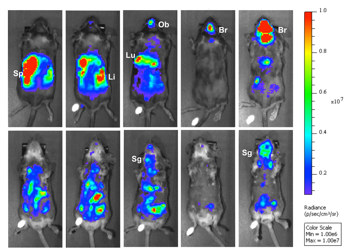
EphrinB2, the receptor for henipaviruses, is present on murine embryonic stem cells (ESC), hematopoietic stem cells (HSC), and neural stem cells (NSC), and has been previously described as a putative molecular marker of “stemness”4. In collaboration with stem cell biologists, we have developed an armamentarium of tools based on the picomolar affinity of NiV-G for EphrinB25-7 to interrogate the role of ephrinB2 in human pluripotent stem cell fate and differentiation. In addition, in collaboration with gene therapists, we are developing Nipah virus Env pseudotyped lentiviral vectors that can cross the blood brain barrier and target the CNS. Our initial efforts are encouraging8 and show that NiV Env pseudotypes can not only target specific populations of human ESC, HSC, and NSC in vitro, but when administered intravenously in vivo, can bypass the liver sink, a critical barrier in targeted gene delivery.